

Aneurysm From Wikipedia, the free encyclopedia.
An aneurysm or aneurism (from Greek: ἀνεύρυσμα - aneurusma "dilation", from ἀνευρύνειν - aneurunein "to dilate") is a localized, blood-filled balloon-like bulge in the wall of a blood vessel. Aneurysms can commonly occur in arteries at the base of the brain (the circle of Willis) and an aortic aneurysm occurs in the main artery carrying blood from the left ventricle of the heart. When the size of an aneurysm increases, there is a significant risk of rupture, resulting in severe hemorrhage, other complications or death. Aneurysms can be hereditary or caused by disease, both of which cause the wall of the blood vessel to weaken.
Classification
Aneurysms may be classified by type, location, and the affected vessel. Other factors may also influence the pathology and diagnosis of aneurysms.True and False Aneurysms
A true aneurysm is one that involves all three layers of the wall of an artery (intima, media and adventitia). True aneurysms include atherosclerotic, syphilitic, and congenital aneurysms, as well as ventricular aneurysms that follow transmural myocardial infarctions (aneurysms that involve all layers of the attenuated wall of the heart are also considered true aneurysms).A false aneurysm or pseudo-aneurysm does not primarily involve such distortion of the vessel. It is a collection of blood leaking completely out of an artery or vein, but confined next to the vessel by the surrounding tissue. This blood-filled cavity will eventually either thrombose (clot) enough to seal the leak or rupture out of the tougher tissue enclosing it and flow freely between layers of other tissues or into looser tissues. Pseudoaneurysms can be caused by trauma that punctures the artery and are a known complication of percutaneous arterial procedures, such as arteriography, arterial grafting, or use of an artery for injection. Like true aneurysms, they may be felt as an abnormal pulsatile mass on palpation.
Morphology
Aneurysms are classified by their macroscopic shape and size and are described as either saccular or fusiform. Saccular aneurysms are spherical in shape and involve only a portion of the vessel wall; they vary in size from 5 to 20 cm (8 in) in diameter, and are often filled, either partially or fully, by thrombus. Fusiform ("spindle-shaped") aneurysms are variable in both their diameter and length; their diameters can extend up to 20 cm (8 in). They often involve large portions of the ascending and transverse aortic arch, the abdominal aorta, or less frequently the iliac arteries. The shape of an aneurysm is not pathognomonic for a specific disease.Location
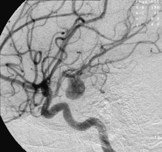
Cerebral aneurysms, also known as intracranial or brain aneurysms, occur most commonly in the anterior cerebral artery, which is part of the circle of Willis. The next most common sites of cerebral aneurysm occurrence are in the internal carotid artery.Many non-intracranial aneurysms arise distal to the origin of the renal arteries at the infrarenal abdominal aorta, a condition some have postulated to be related to atherosclerosis. However, increasing evidence suggests abdominal aortic aneurysms are a wholly separate pathology.
The thoracic aorta can also be involved. One common form of thoracic aortic aneurysm involves widening of the proximal aorta and the aortic root, which leads to aortic insufficiency.
Aneurysms can also occur in the legs, particularly in the deep vessels (e.g., the popliteal vessels in the knee).
Arterial and Venous
Arterial aneurysms are much more common, but venous aneurysms do happen (for example, the popliteal venous aneurysm) ......See the full article Aneurysm From Wikipedia, the free encyclopedia.
Ask a Scientist - Strokes and Aneurysms
Why do people have strokes after having suffered an aneurysm? Researchers answer your questions. For more details check out NRC's online S&T magazine Dimensions.Brain Aneurysm Survivor: John's Story
While exercising on his lunch hour one day, John Davidson felt a sudden, severe shooting pain at back of head: his brain was bleeding. Hear from John and Lori Patsis, RN, about the immediate and life-saving care he received at Northwestern Lake Forest Hospital's Emergency Department.Hemorrhagic Stroke Treatment at Barnes-Jewish Hospital
A hemorrhagic stroke, or what is also called a ruptured brain aneurysm, occurs when the walls of blood vessels are weakened, resulting in a blood-filled bulge. If left untreated, the aneurysm may rupture, causing a hemorrhagic stroke. Immediate and specialized treatment is a key element to minimize the damage, and survival depends on the expertise and speed with which a patient is treated.Debbie was rushed to Barnes-Jewish Hospital after experiencing a hemorrhagic stroke. For more information about stroke treatment, visit http://www.barnesjewish.org/neurosciences/stroke-treatment-rehabilitation.
A hemorrhagic stroke, or what is also called a ruptured brain aneurysm, occurs when the walls of blood vessels are weakened, resulting in a blood-filled bulge. If left untreated, the aneurysm may rupture, causing a hemorrhagic stroke. Immediate and specialized treatment is a key element to minimize the damage, and survival depends on the expertise and speed with which a patient is treated.
The Stroke Team at Barnes-Jewish Hospital knows that time is critical, which is why they have honed treatment down to the second. They treat every stroke patient individually as no two aneurysms are the same, and each patient receives unique treatment based on their symptoms. The specialized critical care unit assigns a team to each patient and supervises the progress to help improve overall outcome.
To learn more, visit http://www.barnesjewish.org/neurosciences/stroke-treatment-rehabilitation.
Brain's Recovery Process After A Stroke or Aneurysm
Rehab specialist Dr. Andrea Toomer of Culicchia Neurological Clinic in New Orleans discusses how cells surrounding the brain recover from the trauma of a stroke or aneurysm. Dr. Toomer was addressing members of the New Orleans Brain Aneurysm Support Group.Brain Aneurysms Animation
Brain Aneurysms, fusiform aneurysms, ruptured aneurysm, coiling of an aneurysm, clipping of an aneurysm.Coil Embolization
This animation shows how a catheter is placed in a cerebral aneurysm, followed by coil placement inside the aneurysm.Neurointerventional Stroke Rounds - Aneurysm Coiling
This 5 minute video walks through treatment of a brain aneurysm by coiling. Dr. Coleman Martin at the Saint Luke's Brain and Stroke Institute in Kansas City, Missouri explains the coiling process with models, animation and angiogram footage of an actual coiling.Mayo Clinic Study on Brain Aneurysms
A new international collaborative study led by Mayo Clinic found that the risk of a brain aneurysm rupturing over time depends on the location and size of the aneurysm.A brain aneurysm, also known as an intracranial or cerebral aneurysm, is an abnormal sac or tiny balloon on a blood vessel to the brain. Aneurysms can rupture and bleed into the area between the brain and the surrounding membrane, leading to stroke and death. An estimated 2 percent of Americans, approximately 6 million people, have brain aneurysms. These aneurysms rupture in about 25,000 people each year.
This study provides us with very useful information that will allow us to better guide our patients with unruptured aneurysms regarding the risk of aneurysm rupture during a very long period of follow-up, says Robert Brown, M.D., a Mayo Clinic neurologist and the studys lead investigator.
This study was part of the International Study of Unruptured Intracranial Aneurysms, which includes 4,059 patients with unruptured brain aneurysms at 61 different medical centers in North America and Europe. These patients have been followed for an average of more than nine years.
Dr. Brown and his team found that rupture risk was somewhat higher among patients with aneurysms in the back of the brain or in the posterior communicating artery, also in the back of the brain, compared to those in the front of the brain. Additionally, patients whose aneurysms were more than 13 millimeters (mm) in diameter were at least twice as likely to experience rupture, compared to those whose aneurysms were 712 mm in diameter.
This study will be presented at the 6th World Stroke Congress meeting in Vienna, Austria, on Sept. 25, 2008.
For more information from Mayo Clinic on aneurysms, click on the following link:
http://www.mayoclinic.org/cerebral-aneurysm/.
No comments:
Post a Comment